Dental Caries: Clinical Overview, Pathophysiology, and Diagnostic Insights
According to the American Dental Association (ADA), a human tooth is composed of three distinct functional layers:
- Enamel: The outermost, highly mineralized acellular protective shield. Lacking living cells, enamel represents the hardest substance in the human body and serves as the primary barrier against mechanical wear and chemical dissolution.
- Dentin: The bulk structure beneath the enamel, containing millions of microscopic dentinal tubules leading directly to the dental nerve plexus. When enamel suffers structural breakdown, exposed dentin transmits thermal (hot and cold), chemical, and tactile stimuli to the nerve, precipitating dentinal hypersensitivity and acute pain.
- Dental Pulp: The neurovascular core located at the center of the tooth. The pulp contains loose connective tissue, blood vessels, lymphatics, and nerves that maintain tooth vitality.
General Overview of Dental Caries
While dental caries is persistent and progressive if left unmanaged, it is fully preventable and manageable in its early stages through effective oral hygiene, diet modification, and remineralization therapies.
What is Dental Caries?
Dental caries develops when acidogenic and aciduric bacteria residing within the dental plaque biofilm metabolize dietary carbohydrates (primarily free sugars). This fermentation process yields organic acids (such as lactic acid) as metabolic waste products, which drastically drop the local pH at the tooth surface below the critical threshold (≈ 5.5).
The prolonged acidic microenvironment induces mineral loss from the hydroxyapatite crystals of the tooth structure. Over time, continuous demineralization outpaces the natural remineralizing capability of saliva, resulting in structural breakdown ranging from superficial enamel white spot lesions to deep dentinal cavitations and endodontic pulp involvement.
Clinical Manifestations of Dental Caries
The clinical presentation of dental caries varies depending on the depth of tissue destruction, ranging from asymptomatic early enamel demineralization to severe endodontic pain.
Primary Signs and Symptoms
Asymptomatic Initial Stage (Enamel Caries): In its earliest phase, demineralization presents visually as a non-cavitated chalky white spot lesion along the gingival margin or pit-and-fissure surfaces, often without any pain or temperature sensitivity.
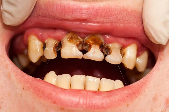
Progressive Enamel and Dentinal Cavitation: As demineralization advances, the white spot breaks down into a visible brown, dark grey, or black structural defect (cavity) on the tooth surface.
Dentinal Hypersensitivity: Once caries breaches the enamel-dentin junction (EDJ), exposed dentinal tubules allow thermal (cold/hot), chemical (sweet/acidic foods), or mechanical stimuli to reach the nerve fibers, triggering sharp, transient pain.
Spontaneous Odontalgia (Toothache): Deep caries extending near the pulp cavity causes continuous, throbbing, or severe toothache that can wake patients from sleep, indicating early or advanced pulpitis (pulp inflammation).
Periodontal and Soft Tissue Involvement: Bacterial spread to surrounding tissues causes localized gingival swelling, tenderness, and pus discharge (suppuration) around the affected tooth.
Odontogenic infections that spread beyond the root apex can result in Ludwig’s Angina-a life-threatening, rapidly progressive cellulitis of the floor of the mouth and submandibular space. This can cause severe soft-tissue swelling, elevation of the tongue, and catastrophic airway compromise requiring emergency surgical drainage and airway management.
Potential Complications of Untreated Dental Caries
Ignoring early caries or dismissing decay in primary (baby) teeth leads to progressive structural breakdown and severe locoregional or systemic complications:
Locoregional Complications
- Irreversible Pulpitis and Apical Periodontitis: Unchecked bacterial invasion leads to irreversible pulpal necrosis and periapical inflammation, causing intense, unrelenting pain.
- Periapical Abscess Formation: Accumulation of purulent exudate at the root apex, manifesting as severe facial edema, fever, localized soft-tissue swelling, and sinus tract drainage.
- Premature Tooth Loss & Malocclusion: Extensive coronal destruction often necessitates extraction. In pediatric patients, premature loss of primary molars leads to loss of arch space, drifting of adjacent teeth, and severe permanent malocclusion.
- Impaired Mastication and Nutritional Deficits: Persistent dental pain hinders proper chewing, forcing individuals to avoid nutrient-dense foods, which can cause significant weight loss or pediatric developmental delays.
Indications for Medical & Dental Evaluation ?
Regular preventive dental checkups are essential because early enamel caries is often completely asymptomatic and reversible with fluoride remineralization therapies.
The pathogenesis of dental caries is driven by the complex interplay between the oral microbiome, host susceptibility, dietary substrates, and time (often conceptualized via the Keyes Triad). Acidogenic Microflora and Biofilm Dynamics: Hundreds of bacterial species naturally inhabit the oral cavity. In a cariogenic environment, acid-producing (acidogenic) and acid-tolerant (aciduric) species-principally Streptococcus mutans, Streptococcus sobrinus, and Lactobacillus species-dominate the dental plaque biofilm. Fermentation of Dietary Carbohydrates: When dietary free sugars (sucrose, glucose, fructose) and refined starches are consumed, plaque bacteria ferment these substrates as an energy source. The metabolic byproducts consist of strong organic acids (primarily lactic, acetic, and propionic acids). The Critical pH Threshold & Critical Demineralization: Salivary Defense Mechanics vs. Demineralization: Saliva serves as the natural primary defense mechanism against tooth decay by: Allowing infants or toddlers to sleep with a bottle containing milk, infant formula, fruit juice, or sugar-sweetened liquids leads to Baby Bottle Tooth Decay. Nocturnal salivary flow drops dramatically during sleep, resulting in prolonged pooling of fermentable carbohydrates around the upper anterior teeth. This causes rapid, rampant demineralization of primary enamel that can destroy the primary dentition within months. Dental caries is a disease of high prevalence across all age brackets, but specific systemic, physiological, and behavioral risk profiles substantially elevate individual susceptibility. Infants and Toddlers (Nursery/Bottle Caries): Young children who are fed sugar-sweetened beverages or milk via nursing bottles prior to naptime or bedtime. Individuals with Hyposalivation or Xerostomia (Dry Mouth): Older Adults and Geriatric Populations: Aging is frequently accompanied by gingival recession, exposing vulnerable root surfaces (cementum and root dentin). Dentin demineralizes at a higher critical pH threshold (≈ 6.2 – 6.7) than enamel, leading to a high incidence of root surface caries. Accurate detection of dental caries requires a combination of clinical visual-tactile inspection, advanced diagnostic imaging, and risk assessment to detect lesions before extensive irreversible structural loss occurs. Visual Examination under High-Intensity Light: Teeth are cleaned and thoroughly dried with compressed air. Clinicians inspect for chalky white spots (incipient enamel caries), micro-cavitations, or dark marginal discolorations using magnification loupes. Tactile Probing with Dental Explorers: Gentle tactile assessment of pits and fissures using a rounded probe (such as an ICDAS-approved ball-ended probe) to feel for soft enamel, surface roughness, or sticky dentin. Standardized Caries Classification Systems: Utilizing frameworks like the ICDAS (International Caries Detection and Assessment System) to categorize lesions from stage 0 (sound tooth surface) to stage 6 (extensive distinct cavity with visible dentin). Forceful probing with sharp, pointed dental explorers into early white spot lesions (non-cavitated enamel caries) is strictly contraindicated. High mechanical pressure can break the fragile, remineralizable outer surface enamel layer, transforming a chemically reversible lesion into an irreversible physical cavitation that requires surgical restoration. Treatment strategy is dictated by the depth of tissue destruction, pulp vitality status, and individual caries risk profile, ranging from non-invasive remineralization to endodontic and surgical therapies. Direct Restorative Materials (Dental Fillings): Indirect Restorations (Inlays, Onlays, and Crowns): Indicated for teeth with extensive coronal structural loss where direct fillings cannot withstand occlusal forces. Adopting structured daily oral hygiene practices and optimizing nutritional habits are foundational to arresting early enamel demineralization, mitigating cariogenic bacterial plaque accumulation, and controlling the progression of active caries. Rigorous Mechanical Plaque Removal: Brush teeth at least twice daily for two minutes using a soft-bristled toothbrush. Employing the Modified Bass Technique (angling bristles at 45°C toward the gingival margin with gentle circular motion) ensures thorough plaque removal along the cervical margins and interproximal spaces. Interdental Cleansing Protocols: Traditional toothbrushing cleans only approximately 60% of tooth surfaces. Daily utilization of dental floss, interdental brushes, or oral irrigators (water flossers) is mandatory to disrupt interproximal plaque biofilms where proximal caries most frequently originate. Systemic and Topical Hydration: Maintain a high intake of plain fluoridated water (2 to 2.5 liters daily) to prevent xerostomia (dry mouth), encourage continuous salivary flow, and enhance mechanical clearance of residual food particles. Dietary Sugar Regulation & Frequency Control: Adherence to Professional Dental Recalls: Schedule routine dental checkups every 6 months for professional prophylaxis (scaling and polishing) and early detection of incipient non-cavitated lesions. Avoid brushing teeth immediately after consuming highly acidic foods or beverages (such as citrus fruits, soda, or wine). Direct mechanical scrubbing when enamel is temporarily softened by acid accelerates enamel wear and loss. Patients should wait at least 30 to 60 minutes after acidic intake-or rinse thoroughly with plain water or a bicarbonate solution-before brushing to allow natural salivary remineralization to re-harden the enamel matrix. Preventing dental caries requires a comprehensive approach leveraging mechanical biofilm control, targeted chemical remineralization, and dietary management: Optimal Topical Fluoride Therapy: Application of Pit and Fissure Sealants: Application of flowable resin or glass-ionomer sealants over the deep, narrow occlusal grooves of permanent molars in children and adolescents. Sealants form a physical barrier, isolating vulnerable tooth anatomy from acidogenic bacteria and food debris. Salivary Stimulation and Xylitol Usage: Chewing sugar-free gum containing Xylitol after meals stimulates protective salivary flow while inhibiting the metabolic growth of Streptococcus mutans, reducing overall cariogenic plaque accumulation. Tobacco Cessation and Substance Management: Eliminate tobacco smoking, vaping, and excessive alcohol consumption, as these habits diminish salivary flow, alter oral microbial ecology, and accelerate periodontal tissue breakdown.Patients should seek an immediate evaluation by a dental professional if:
Primary Etiologies and Pathophysiology
Major Etiological Mechanisms:

Epidemiological Risk Factors
High-Risk Demographics ?
Aggravating Risk Factors
Diagnostic Methodologies for Dental Caries
Clinical and Visual-Tactile Inspection
Radiographic and Advanced Diagnostic Modalities
Evidence-Based Clinical Management Strategies
Non-Invasive Remineralization Therapy (Early/Incipient Caries)
Operative Restorative Dentistry (Cavitated Caries)
Advanced Endodontic and Surgical Interventions
Lifestyle Modifications and Home Management
Proactive Prevention Strategies