
Otitis Media with Effusion: Clinical Overview, Pathophysiology, and Symptoms
General Overview of Otitis Media with Effusion
OME is recognized as the leading cause of acquired hearing loss in early childhood. It frequently develops following an episode of acute otitis media (AOM) or secondary to Eustachian tube dysfunction. Chronic OME is diagnosed when fluid persistence exceeds 3 consecutive months from the time of onset or clinical detection.
What is Otitis Media with Effusion?
Otitis media with effusion occurs when fluid accumulates in the tympanic cavity and Eustachian tube. This persistent fluid dampens the mechanical mobility of the tympanic membrane (eardrum) and ossicular chain, obstructing normal acoustic wave conduction to the inner ear and resulting in conductive hearing loss.
While OME lacks active bacterial suppuration, the presence of stagnant fluid alters the local tissue environment, predisposing the middle ear to recurrent secondary infections.
Signs and Symptoms of Otitis Media with Effusion
Clinical Manifestations
- Conductive Hearing Loss: The hallmark sign, often presenting as mild to moderate hearing reduction or muffled sound perception.
- Balance Impairment and Equilibrium Issues: Fluid-induced pressure changes within the middle ear can disrupt vestibular function, leading to clumsy gait or unsteadiness.
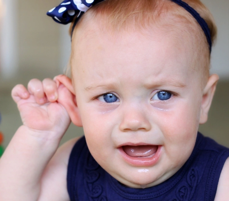
- Pinna or Auricular Deformities: Secondary structural changes or discomfort causing children to pull or tug at the outer ear.
- Developmental and Speech Delays: In young children, persistent unrecognized hearing deficit can delay speech and language acquisition.
- Behavioral Indicators: Frequent crying, irritability, pulling at ears, and unexplained restlessness, especially in non-verbal pediatric patients.
Health Impact of OME
OME presents significant challenges, particularly in pediatric populations:
- In Adults and Older Children: Persistent ear fullness, mild otalgia, decreased auditory acuity, and reduced academic or occupational performance.
- In Infants and Toddlers: Inability to articulate discomfort manifests as persistent crying, sleep disturbances, and parental distress regarding delayed speech progression.
Potential Complications of Untreated OME
When OME persists or recurs frequently, it can trigger severe structural and regional sequelae:
- Tympanic Membrane Perforation: Chronic middle ear fluid pressure can compromise eardrum integrity, leading to persistent perforation, abnormal ear discharge, and chronic hearing deficit.
- Mastoiditis: Spread of inflammation to the air cells of the mastoid bone, presenting with retroauricular swelling, erythema, tenderness, and external auditory canal displacement.
- Middle Ear Cholesteatoma & Scarring: Formation of benign keratinizing epidermal cysts in the middle ear space or tympanosclerosis (scarring and hyalinization of the eardrum).
- Recurrent Acute Otitis Media: Stagnant fluid acts as a culture medium for opportunistic pathogens, fueling frequent acute infectious flare-ups.
Indications for Immediate Medical Evaluation
Early otolaryngologic assessment is essential to preserve hearing function and prevent chronic structural damage.

Primary Etiologies and Pathophysiology of OME
Otitis media with effusion develops primarily from persistent Eustachian tube dysfunction (ETD) or incomplete resolution of acute middle ear inflammation, leading to fluid accumulation without active suppuration.
Key Pathological Mechanisms:
Prior Acute Otitis Media: Following an episode of acute bacterial or viral otitis media, active infection clears, but inflammatory exudate and serous fluid often remain trapped within the middle ear cavity. This residual fluid can persist for weeks or months, transitioning into chronic OME.
Eustachian Tube Dysfunction & Mechanical Obstruction: The Eustachian tube regulates middle ear gas pressure and drains physiological secretions into the nasopharynx. Functional or anatomical blockages-such as hypertrophied adenoids, nasopharyngeal tumors, or improper feeding postures (e.g., bottle-feeding while lying flat)-impair tube opening. This creates persistent negative pressure within the tympanic cavity, pulling transudate fluid from mucosal capillaries into the middle ear space.
Allergic Reactions and Hypersensitivity: Inhaled environmental allergens (such as dust mites, pollen, pet dander, or secondhand tobacco smoke) trigger local mucosal allergic responses. Mucosal edema within the nasopharynx and Eustachian tube orifice hinders normal fluid drainage and increases middle ear secretory activity.
Sudden Barometric Pressure Fluctuations: Rapid ambient pressure shifts (barotrauma)-experienced during air travel descent, scuba diving, or driving through high-altitude mountain passes-can cause functional closure of the Eustachian tube. The resulting pressure differential injures the middle ear mucosa and promotes fluid accumulation.
Epidemiological Risk Factors for OME
High-Risk Demographics?
Infants and Young Children (Aged 6 Months to 3 Years): Pediatric patients are highly susceptible due to their immature immune systems and shorter, wider, and more horizontal Eustachian tubes, which facilitate fluid stagnation and reflux from the nasopharynx.
Children Attending Group Daycare: High exposure to viral upper respiratory infections significantly increases the incidence of Eustachian tube inflammation and OME.
Individuals with Craniofacial Abnormalities: Children diagnosed with cleft palate, Down syndrome, or muscular dysfunctions that affect the tensor veli palatini muscle experience chronic Eustachian tube dysfunction.
Patients with Untreated Upper Respiratory Allergies: Individuals with chronic allergic rhinitis or adenoid hypertrophy.
Aggravating Risk Factors
- Passive Tobacco Smoke Exposure: Secondhand smoke impairs ciliary function within the respiratory epithelium, increasing mucus viscosity and Eustachian tube inflammation.
- Bottle-Feeding in a Supine Position: Feeding infants while they lie flat allows milk or formula to enter the Eustachian tube orifice, inducing local inflammation and fluid buildup.
- Pacifier Use Beyond Early Infancy: Prolonged or frequent pacifier use alters swallow mechanics and pressure dynamics within the nasopharynx, predisposing young children to Eustachian tube dysfunction.
- Seasonality: A higher prevalence is observed during autumn and winter months, coinciding with peak incidence of viral upper respiratory tract infections.
Diagnostic Methodologies for OME
Establishing a definitive diagnosis of otitis media with effusion requires differentiating fluid buildup from acute suppurative otitis media and evaluating the degree of auditory impairment.
Clinical Evaluation & Risk Profile Assessment
Clinical Presentation: Clinicians evaluate key signs such as fluctuating conductive hearing impairment, a sense of aural fullness, autophony (hearing one’s own voice echo), balance instability, or speech/language delays in pediatric patients without systemic infection signs (such as high fever or severe otalgia).
Risk Factor Screening: Reviewing predisposing patient background, including recent upper respiratory tract infections (URTIs), history of acute otitis media, perennial allergic rhinitis, craniofacial anomalies (e.g., cleft palate), or passive exposure to environmental tobacco smoke.
Objective Otologic Examination Techniques
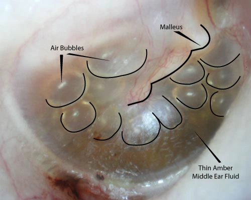
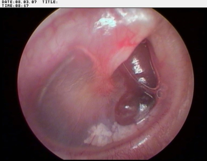
Pneumatic Otoscopy: The primary clinical standard for diagnosing OME. Visual inspection of the tympanic membrane (eardrum) typically reveals:
- An intact, opaque, dull, or amber/bluish eardrum.
- Visible air-fluid levels or intratympanic air bubbles behind the membrane.
- Retraction or loss of normal eardrum translucency and bony landmarks.
- Significantly impaired or absent tympanic membrane mobility when positive and negative air pressure is applied via the pneumatic bulb.
Tympanometry: An objective electroacoustic test that measures middle ear compliance and air pressure dynamics:
- Type B Tympanogram (Flat curve): Indicates fluid accumulation within the tympanic cavity with restricted eardrum compliance, strongly confirming OME.
- Type C Tympanogram: Demonstrates significant negative pressure within the middle ear space, reflecting advanced Eustachian tube dysfunction.
Audiometric Testing: Pure-tone audiometry (or behavioral observation audiometry in infants) is performed to measure the baseline conductive hearing loss threshold and assess its functional impact on verbal communication.
Evidence-Based Clinical Management Strategies
Therapeutic management of OME aims to clear middle ear effusion, restore normal atmospheric pressure, recover hearing acuity, and prevent long-term structural sequelae.
Pharmacological Therapy
In uncompromised patients without developmental risks, an initial period of watchful waiting for 3 months is widely recommended, as many cases of post-infectious OME resolve spontaneously.
- Targeted Symptomatic & Allergy Management: Antihistamines, intranasal corticosteroids, or leukotriene receptor antagonists may be prescribed if OME is directly driven by allergic rhinitis or upper airway mucosal inflammation.
- Avoidance of Ineffective Regimens: Clinical practice guidelines generally advise against routine long-term use of systemic antibiotics, oral decongestants, or systemic steroids solely for OME, as they fail to demonstrate long-term efficacy in clearing chronic fluid.
Surgical Interventions
Surgical management is indicated when effusion persists for more than 3 to 6 months, or when chronic OME causes significant structural drum changes, persistent conductive hearing loss (≥20-30dB), or developmental speech delays.
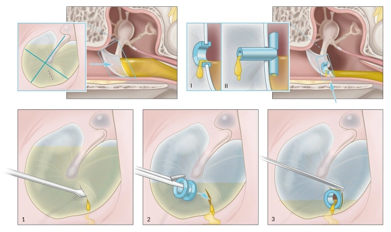
- Myringotomy with Tympanostomy Tube Insertion: A micro-incision is made in the tympanic membrane to aspirate trapped fluid, and a tiny grommet/tube is placed. This restores ventilation, equalizes middle ear pressure, and lowers the risk of fluid re-accumulation.
- Adenoidectomy: Often performed alongside tympanostomy tube insertion in children with chronic adenoid hypertrophy or persistent nasopharyngeal inflammation obstructing the Eustachian tube orifice.
Acoustic Amplification & Non-Surgical Assistive Devices
- Hearing Aids: Prescribed for pediatric or adult patients with persistent OME who are poor surgical candidates, decline invasive procedures, or continue to experience significant bilateral conductive hearing loss despite surgical intervention.
- Autoinflation Devices: Mechanical devices (such as specialized nasal balloons) that elevated nasal pressure during swallowing to help force air into the Eustachian tube, aiding middle ear ventilation.
Lifestyle Modifications and Home Care Habits
Adopting proactive daily habits and following targeted clinical guidance can manage the progression of otitis media with effusion, prevent secondary middle ear infections, and support natural Eustachian tube drainage.
- Strict Clinical Adherence and Follow-Up: Consistently follow the prescribed treatment plan from your otolaryngologist and attend scheduled follow-up visits to monitor fluid absorption and hearing thresholds.
- Prompt Medical Consultation: Seek immediate medical evaluation if new or worsening symptoms emerge, such as severe otalgia, fever, purulent ear discharge, or sudden hearing drop.
- Proper Nasal and Hygiene Care: Practice gentle nasal hygiene using isotonic saline sprays to clear nasopharyngeal secretions. Avoid forceful blowing of both nostrils simultaneously, as high pressure can force nasal fluid back into the Eustachian tube orifice.
- Nutritional and Immune Support: Maintain a well-balanced, nutrient-dense diet and engage in age-appropriate physical activity to strengthen systemic immunity and lower the incidence of viral upper respiratory infections.
- Correction of Anatomical and Airway Obstructions: Consult an ENT specialist to evaluate and manage underlying anatomical issues that hinder ventilation, such as hypertrophied adenoids, chronic sinusitis, or nasal septal deviation.
Proactive Prevention Strategies
Preventing otitis media with effusion focuses on protecting Eustachian tube function, limiting exposure to environmental irritants, and reducing viral transmission:
- Avoid Supine Infant Feeding: Ensure infants are fed in an upright or semi-inclined position. Avoid feeding babies while they lie flat on their backs to prevent liquid or formula from refluxing into the Eustachian tubes.
- Eliminate Environmental Tobacco Smoke: Protect infants and children from secondhand tobacco smoke and air pollutants, which irritate the respiratory mucosal lining, impair ciliary movement, and cause Eustachian tube edema.
- Prevent Upper Respiratory Infections: Practice frequent handwashing with soap and water, avoid close contact with individuals suffering from acute colds or flu, and maintain routine pediatric vaccinations (including pneumococcal conjugate and annual influenza vaccines).
- Limit Pacifier Duration: Reduce or discontinue pacifier use after 6 months of age, as repetitive sucking mechanics can alter nasopharyngeal pressure dynamics and impair Eustachian tube drainage.
- Manage Underlying Allergies: Treat allergic rhinitis effectively using allergen avoidance techniques or targeted allergy therapy to prevent secondary middle ear mucosal swelling.